In Conversation with Dr Bejoy Thomas
Tuning in to a Different Frequency
Inspirations & Lessons
SPIN : What drew you to radiology, and then specifically to pediatric neuroimaging, at a time when it was barely recognized as its own subspecialty in India?
BT: I trained as a medical student in the late 1980s, a time when diagnostic radiological investigations were primarily limited to X-ray radiography. During my third year of MBBS, an ultrasound machine was introduced to the ‘X-ray department’. Our Head of Radiology, Prof. K. J. Joseph, was incredibly passionate about teaching us young undergraduates about the enormous potential of this emerging field. I remember him showing us MRI pictures of the brain from an atlas; it instantly captured my fascination.
I went on to pursue an MD in Radiodiagnosis for my postgraduate studies. Two primary aspects drew me to radiology: pathological anatomy and the physics of imaging.
Immediately following my MD, I completed a brief stint at a brain and spine hospital, which I found deeply intriguing. That same year, I was fortunate enough to be selected for a fellowship in Neuro and Cardiovascular Radiology at SCTIMST in Thiruvananthapuram, Kerala, India. At the time, this was one of only two fellowship positions available in this field across the entire country.
Years later, following my advanced neuroimaging fellowship in KU Leuven Belgium, I was offered another opportunity to pursue a clinical fellowship in Pediatric Neuroradiology at the prestigious Hospital for Sick Children (SickKids) in Toronto. This experience truly transformed me into a pediatric neuroradiologist. Currently, I practice both adult and pediatric diagnostic neuroimaging as a faculty member at SCTIMST.
SPIN: Your training took you from Kottayam to Trivandrum, then to Leuven as a BOYSCAST scholar, and later to the Hospital for Sick Children in Toronto. How did each of these stops shape the neuroradiologist you became?
BT: I completed my undergraduate training at Government Medical College, Kottayam, finishing my MBBS with the second rank in the university. Later, after being selected through the All India Postgraduate Entrance Examination, I earned my MD from NHL Municipal Medical College in Ahmedabad, graduating with the university's first rank.
Because radiology seats were extremely limited, I had to travel far to pursue my dream, but it turned out to be a remarkable experience. I subsequently had the opportunity to complete my postdoctoral certification (fellowship) in Trivandrum and then stayed on as a faculty member in the Department of Imaging Sciences and Interventional Radiology at SCTIMST. During my tenure as faculty here, a Government of India grant called the BOYSCAST fellowship allowed me to pursue my first advanced neuroimaging fellowship abroad in Belgium, working alongside Dr. Stefan Sunaert and Prof. Paul van Hecke. Professor Sunaert was instrumental in shaping me as a neuroimaging researcher.
Upon returning home, we developed one of the first clinical functional MRI programs in the country.Alongside Prof. C. Kesavadas and many of my colleagues, I was able to publish extensively in the field of advanced neuroimaging, primarily focusing on adult neuroimaging.
A few years later, I was fortunate to be selected for a fellowship in Pediatric Neuroimaging at the prestigious Hospital for Sick Children (SickKids) in Toronto. There, I had the opportunity to train under Dr. Susan Blaser, Dr. Charles Raybaud, and Dr. Manohar Shroff—just to name a few giants in the field. That was one of the most rigorous, yet equally fascinating, periods of my medical training.
Looking back, I feel blessed to have been in the right place at the right time. I am incredibly proud and happy that, after completing my training in the West, I was able to return home to contribute to the field and help train the next generation of great minds in neuroradiology, both in India and abroad.
Innovations & AI
SPIN: What first drew you to quantitative techniques (SynMRI, ASL) over purely qualitative reads?
BT: I have always been fascinated by emerging MRI technologies and their applications in neuroimaging. My initial interest was in Susceptibility Weighted Imaging (SWI), which later shifted to perfusion imaging. From there, it was a natural progression to explore non-invasive perfusion techniques and quantitative methods in neuroimaging.
One of my first major publications—co-authored with Prof. Stefan Sunaert—focused on quantitative Diffusion Tensor Imaging (DTI) in pediatric neuroimaging. It was published in the journal Brain and was featured on the cover of their November 2005 issue (https://doi.org/10.1093/brain/awh600). Ever since that milestone, we have been firmly convinced of the need for quantification in the imaging sciences, a focus we continued to pursue and develop upon returning home.
SPIN: How do you see AI and quantitative imaging converging in the next decade?
BT: Artificial intelligence represents both a boon and a threat to the future of neurological research. Next-generation foundational models and Artificial General Intelligence (AGI) have the potential to make quantitative MR research easier, more reliable, and more reproducible. However, we must remain highly cautious regarding their interpretation in clinical settings until robust levels of explainability are incorporated into these "black box" workflows.
SPIN: What do you think are the biggest barriers to real-world AI adoption in pediatric neuroradiology, particularly in resource-varied settings like India?
BT: The significant variability of normative data across different pediatric age groups, coupled with a relative scarcity of large datasets, presents unique challenges. While generative networks may help overcome these issues to a certain extent, I believe this progression will take much longer in pediatric neuroimaging compared to adult studies. Consequently, this will pose an even greater challenge in resource-limited settings.
Wisdom From a Pioneer
SPIN: You've been central to SCTIMST's epilepsy care programme, which brought together neurologists, neurosurgeons, and neuroradiologists and won the BMJ South Asia Medical Team award. What made that interdisciplinary model work?
BT: Prof. K. Radhakrishnan, a renowned epileptologist at our institute, invited us to join the Epilepsy Care program as neuroradiologists at a time when interdisciplinary decision-making processes were rare in the country. This provided us with a fantastic opportunity to understand the pathophysiology of epilepsy and the critical need for dedicated imaging in the management of such disorders. Later, we initiated multidisciplinary meetings focusing on neuroradiology-pathology and pediatric neuroimaging -genetic correlations, which ultimately helped train an entire generation of neuroradiologists better.
SPIN: How do you approach building bridges between institutions and countries?
BT: This is a very important aspect of one's professional development. Over the years, I have been fortunate enough to maintain strong relationships with my former professors and many friends in the field, both within India and abroad. Thanks to these connections, several of our former trainees have had the opportunity to pursue fellowships at these international institutions, eventually going on to become consultants at various universities around the globe.
SPIN: If you could change one thing about how pediatric neuroimaging training or practice works in India today, what would it be and why?
BT: India needs more specialist-trained pediatric neuroradiologists, as it is a completely different ball game. The best way to improve training is to create more fellowship opportunities within the country for this subspecialty.
SPIN: You've mentored and co-authored with a wide network of trainees over the years. What do you look for when you take someone on as a mentee?
BT: One of the greatest blessings in my life has been the opportunity to work with brilliant minds. In my opinion, the most essential qualities are dedication to the field and the ability to think and adapt to the highest standards of neuroimaging, even within resource-limited settings.
Personal Reflections
SPIN: Is there a case from your career—perhaps a child whose imaging changed the course of their care—that still stays with you?
BT: Many years ago, a young girl came to our hospital presenting with a rubral tremor. This was caused by central brain herniation secondary to intracranial hypotension. Her imaging immediately pointed to the correct diagnosis—an uncommon clinical presentation of Spontaneous Intracranial Hypotension (SIH)—which allowed her to receive the appropriate treatment promptly. I have encountered several such examples that have brought me immense satisfaction, alongside a few disappointments as well.
SPIN: How do you balance a demanding academic and clinical career with the other things that bring you joy?
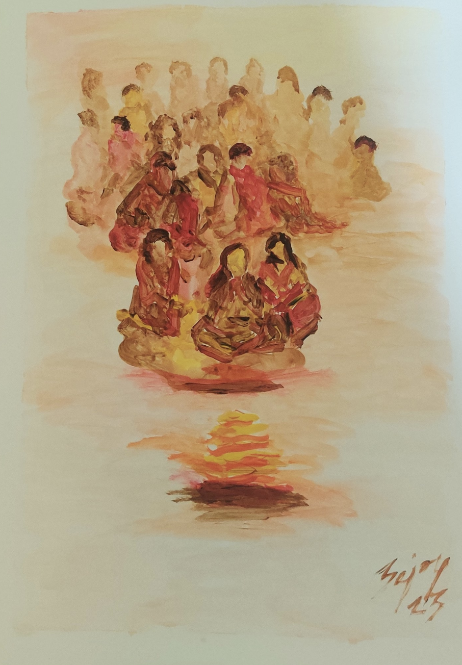
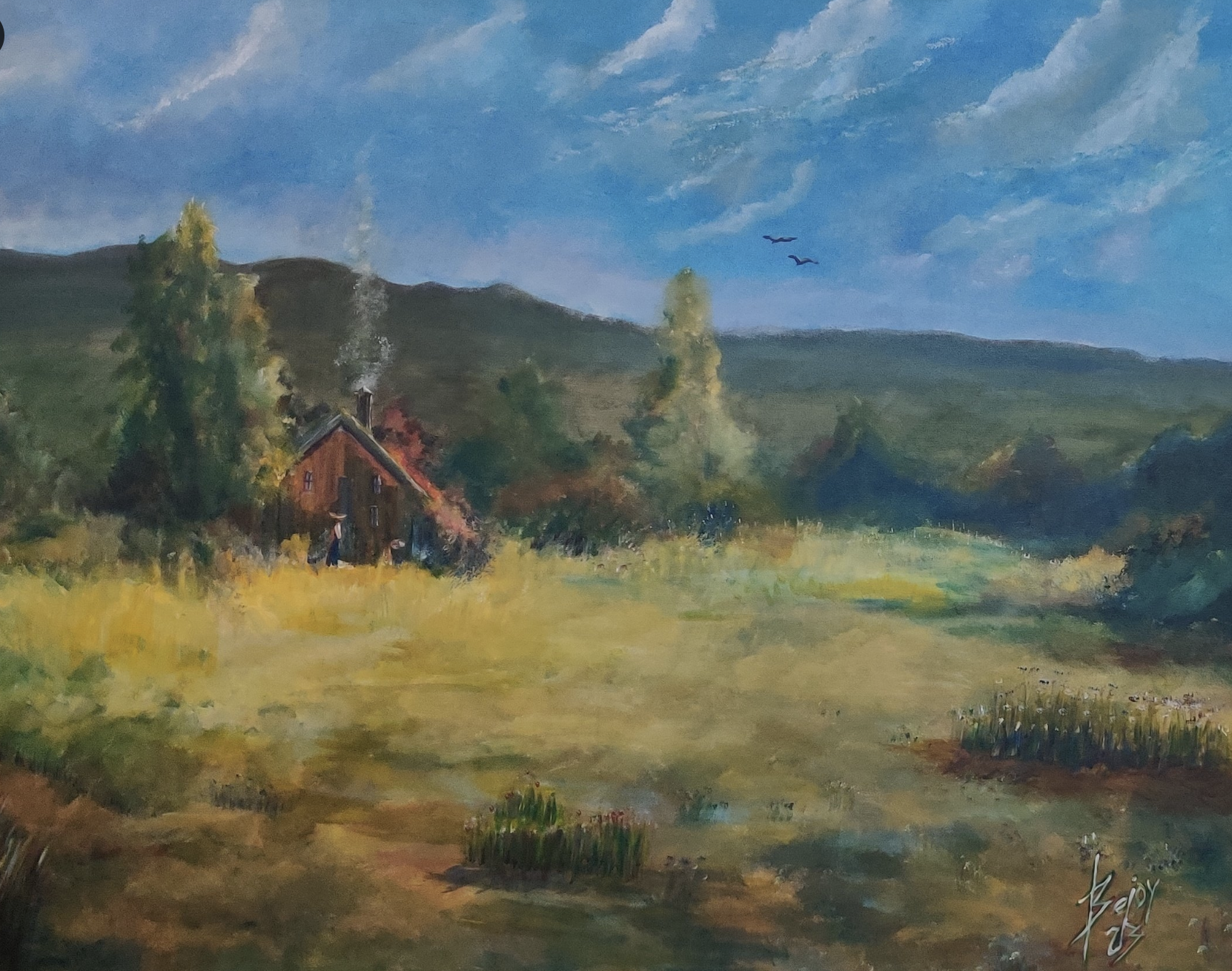
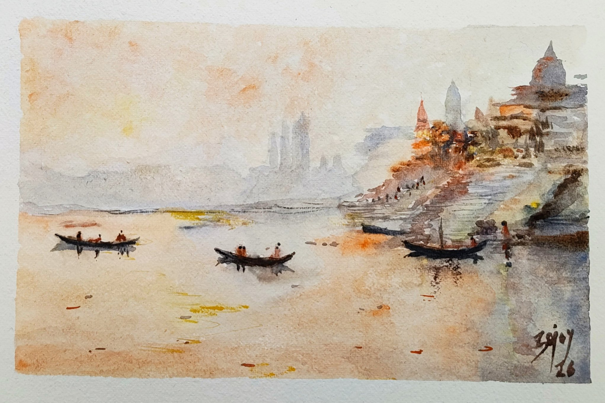
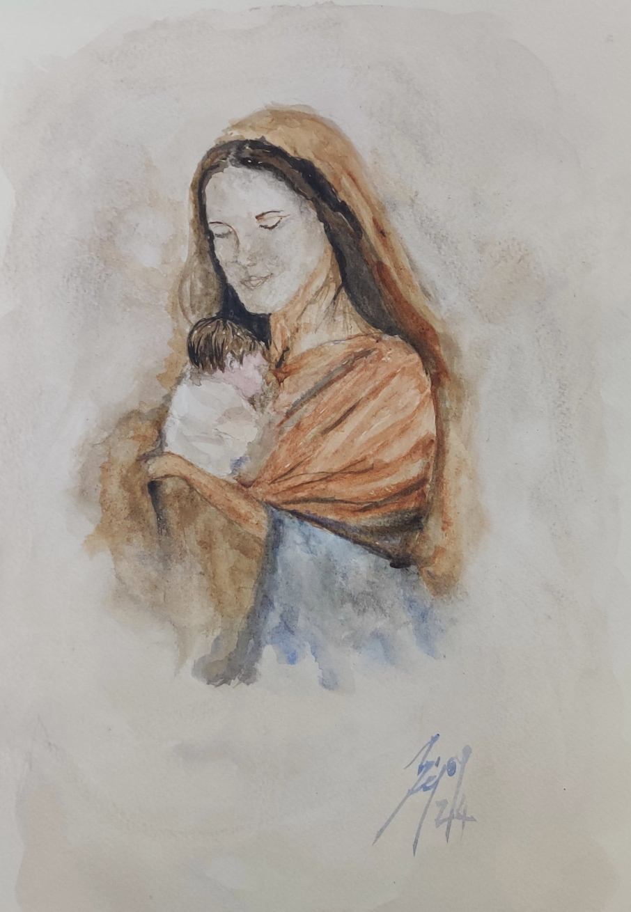
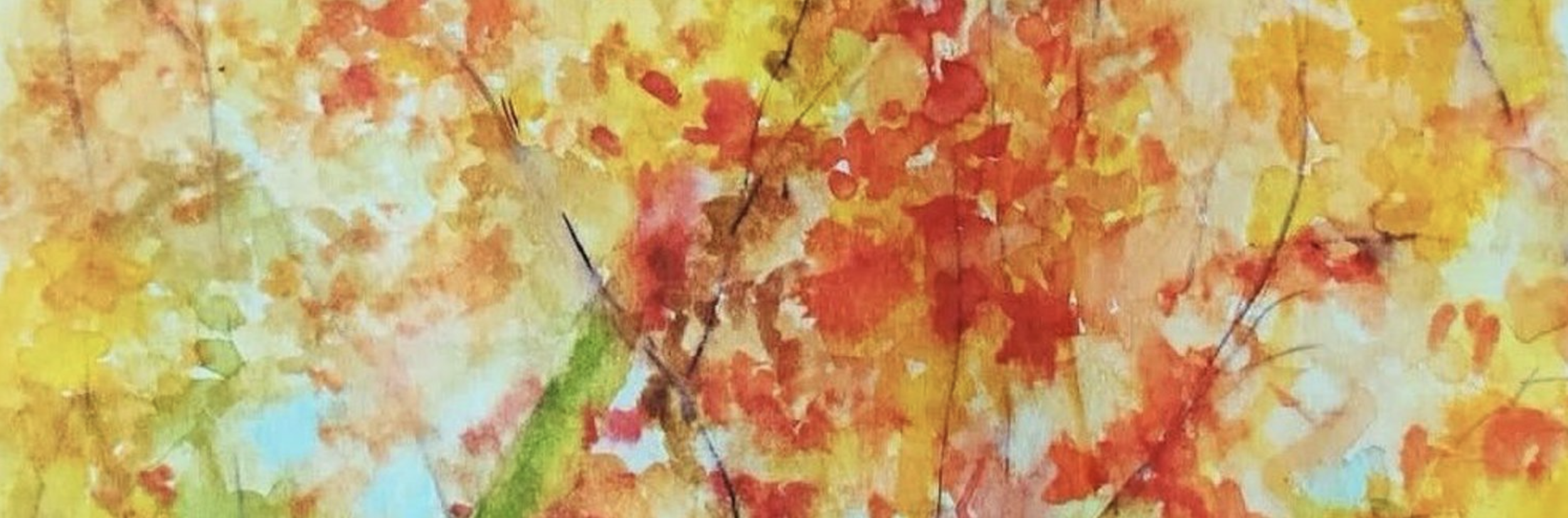
BT: I enjoy painting and singing. As a watercolor and acrylic artist, painting provides me with immense joy and a deep sense of relaxation.
Dr. Bejoy Thomas, MD, DNB, PDCC
Fellowship in Advanced Neuroimaging, KU Leuven, Belgium,
Clinical Fellowship in Pediatric Neuroradiology, The Hospital for Sick Children, U of T, Toronto, Canada

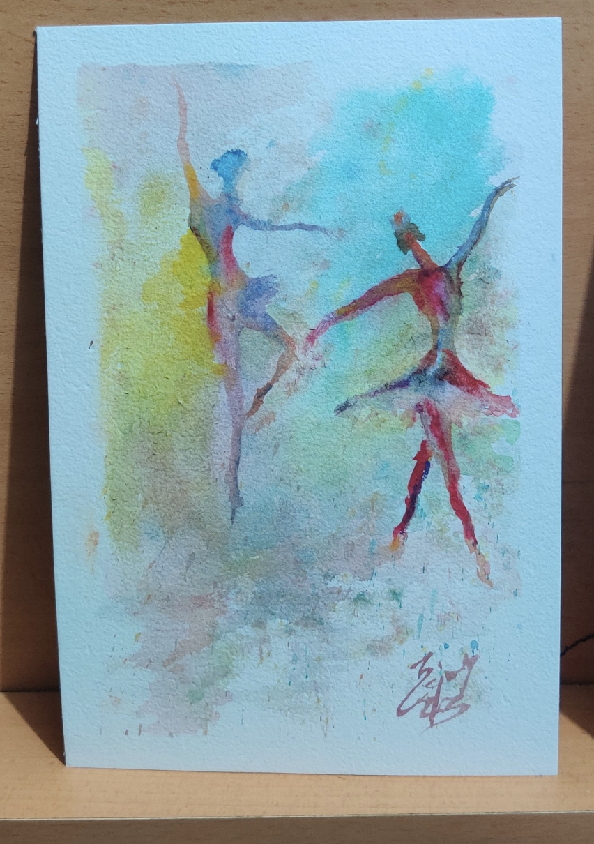
"Dance- pontine tegmental cap and Ponto cerebellar dysplasia "